
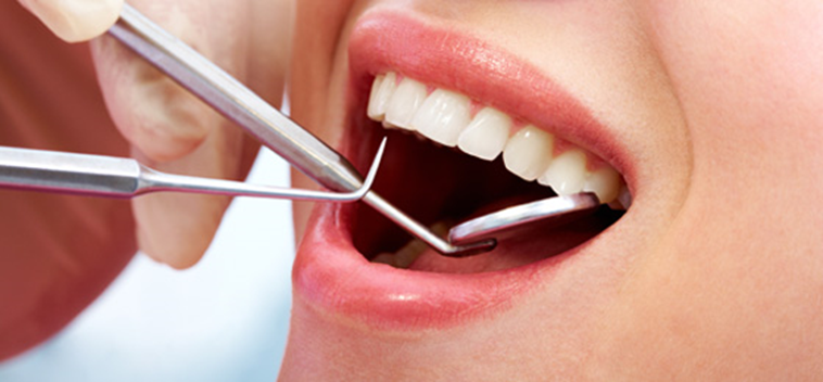
AESTHETIC DENTISTRY:
PROCEDURES PERFORMED :
- Smile Designing
- Bonding: Bonding is a procedure where a small part of composite material is directly applied and sculpted and finally cured to the desired shape.
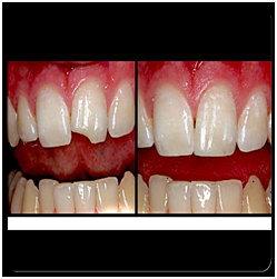
- Porcelain Veneers: Veneers are ultra thin laminates that can be attached directly to the tooth, using a dual cured resin based cement. Used to close gaps between teeth and in some cases may require minimal tooth preparation, for achieving aesthetics.
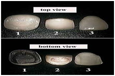
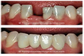
CROWN & VENEERS
CROWNS AND BRIDGES:
Full Coverage Restorations
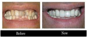
Single tooth replacement
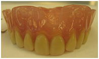
DENTURES:Also known as false teeth are prosthesis constructed to replace missing teeth, which are supported by the surrounding soft & hard tissues of the oral cavity:
REMOVABLE PROSTHODONTICS:
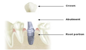
IMPLANT DENTISTRY
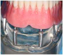
FIXED PROSTHODONTICS
A Dental Implant is an artificial replacement of the root of a missing tooth, which can serve as an abutment for crown, bridge or an over denture.
FIXED REMOVABLE PROSTHODONTICS
ENDODONTICS:
WHY?
To remove Infection from the root canal space of the tooth, which houses the nerve and blood vessels of the tooth.
Due to long standing trauma, the tooth nerve can get necrosed long before the patient notices.
Steps In Root Canal Treatment:
- Access preparation
- Working Length Determination
- Bio Mechanical Preparation
- Obturation
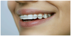
ORTHODONTICS
To improve Aesthetics and function.
Indications:
- Crowding
- Increased Over jet
- Increased traumatic Overbite.
- Anterior Cross Bites (where there is decrease in labial periodontal support of affected lower incisors).
- Un-erupted Impacted Teeth( where there is a danger of Pathology)
- Cross bites associated with Mandibular Displacement
FIXED ORTHODONTIC APPLIANCE
REMOVABLE ORTHODONTIC APPLIANCE
Periodontics:
To Remove Plaque. Enamel Dissolution Occurs in Caries caused by Organic Acids produced by Plaque Bacteria
Majority of people in modern commercialized societies consume a soft sugar- rich diet, which provides for extensive bacterial fermentation, LEADING TO CARIES, as a result of under use of the muscles of mastication, and decreased salivary flow and less stimulation of the tissues.
It also causes Atrophy of the Periodontium which may lead to periodontal disease.
Plaque removal:
Non Surgical:
Scaling and Root Planing
1>
Air Drying shows the supra gingival deposits, which otherwise goes unnoticed
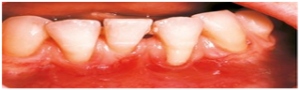
Surgical Removal Of Plaque:
Periodontal Flap Surgery following Scaling and Root Planing.
- Bone grafts. This procedure involves using fragments of your own bone, synthetic bone, or donated bone to replace bone destroyed by gum disease. The grafts serve as a platform for the regrowth of bone, which restores stability to teeth.
Soft tissue grafts. This procedure reinforces thin gums or fills in places where gums have receded.
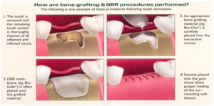
THE SUTURES PLACED AFTER PERIODONTAL FLAP SURGERY
ORAL SURGERY
EXTRACTION:
EXTRACTION OF IMPACTED THIRD MOLAR.
POST-EXTRACTION INSTRUCTIONS:
- BITE ON A PIECE OF SURGICAL GAUZE FOR 30-45 MIN, FOLLOWING THE EXTRACTION, FOR THE FORMATION OF THE CLOT, WHICH IN TURN WILL AID IN THE HEALING OF THE WOUND.
- AVOID SPITTING, VIGOROUS RINSING, BRUSHING NEXT TO THE SITE OF EXTRACTION FOR THE NEXT 72 HOURS.
- DRINK PLENTY OF FLUIDS AND HAVE A SOFT DIET. AVOID HOT LIQUIDS AND FOOD.
- IN CASE OF SWELLING, APPLY AN ICE PACK, OR TAKE FEW ICE CUBES, WRAPPED IN A TOWEL AND APPLY TO THE CHEEK NEXT TO THE EXTRACTION SITE, INTERMITTENTLY FOR THE NEXT 36 HOURS. THE SWELLING WILL COME DOWN AFTER 48 HOURS.
- AVOID SMOKING, ALCOHOL AND ANY FORM OF VIGOROUS EXERCISE IMMEDIATELY AFTER THE EXTRACTION.
- IN CASE OF SECONDARY HEMMORHAGE BITE TIGHTLY ON A PIECE OF SURGICAL COTTON OR GAUZE OR EVEN A CLOTH FOR 30MIN.
- YOUR SURGEON WILL REMOVE THE SUTURES AFTER A WEEK.
Tooth Extraction Recovery
In cases of simple tooth extraction, recovery occurs in 7-10 days whereas in case of surgical extraction, it may take 3 weeks to 3 months for recovery to take place depending on the degree of damage to the dental tissues. In cases of simple extraction, healing after 7-10 days is good enough that a person can eat hard and crusted food without any pain or discomfort. Healing in the oral cavity is faster as compared to the skin because of rich blood supply of the area. If a cut is there on the skin, then it takes longer time to heal than a cut in the oral cavity.
ORAL RADIOLOGY
INTRA-ORAL:
This preoperative photo of tooth #3, (A), reveals no clinically apparent decay other than a small spot within the central fossa. In fact, decay could not be detected with an explorer. Radiographic evaluation, (B), however, revealed an extensive region of demineralization within the dentin (arrows) of the mesial half of the tooth. When a bur was used to remove the occlusalenamel overlying the decay, (C), a large hollow was found within the crown and it was discovered that a hole in the side of the tooth large enough to allow the tip of the explorer to pass was contiguous with this hollow. After all of the decay had been removed, (D), the pulp chamber had been exposed and most of the mesial half of the crown was either missing or poorly supported.
IOPA: INTRA-ORAL PERI-APICAL
Intra-oral Peri-apical Radiographic views allow:
- Detection of Dental Cavities
- Pulpal pathology
- Study the Roots
- Periodontal Pathology
- Periapical Infection.
BITEWING:
Ø -small areas of decay between the teeth or below existing restorations (fillings)
Ø -infections in the bone
Ø -periodontal (gum) disease
Ø -abscesses or cysts
Ø -developmental abnormalities and some tumors.
EXTRA-ORAL:
- OPG:Impacted wisdom teeth diagnosis and treatment planning – the most common use is to determine the status of wisdom teeth and trauma to the jaws.
- Periodontalbone loss and periapical involvement.
- Finding the source of dental pain
- Assessment for the placement of dental implants
- Orthodontic pre and post operative
- Diagnosis of developmental anomalies such as cherubism,cleido cranial dysplasia
- Carcinoma in relation to the jaws
- Diagnosis of osteosarcoma, ameloblastoma, renal osteodystrophy affecting jaws and hypophosphatemia.
- Diagnosis, and pre- and post-surgical assessment of oral and maxillofacial trauma, e.g. dentoalveolar fractures and mandibular fracture
- Salivary stones (Sialolithiasis).
- Other diagnostic and treatment applications.[1]
- CBCT
IMPORTANCE OF CONE BEAM CONE TOMOGRAPHY:
Dental cone beam CT is commonly used for treatment planning of orthodontic issues. It is also useful for more complex cases that involve:
- surgical planning for impacted
- diagnosing temporomandibular joint disorder (TMJ).
- accurate placement of dental implants.
- evaluation of the jaw, sinuses, nerve canals and nasal cavity.
- detecting, measuring and treating jaw tumors.
- determining bone structure and tooth orientation.
- locating the origin of pain or pathology.
- cephalometric analysis &reconstructive surgery